|
Contec medical systems Co., Ltd
|
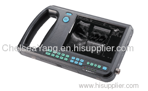
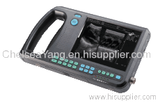
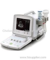
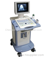
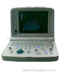
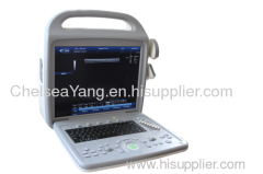
Digital PalmSmart Uitrasound Scanner
| Payment Terms: | WU;Paypal or Wire Transfer |
| Place of Origin: | Hebei, China (Mainland) |
|
|
|
| Add to My Favorites | |
| HiSupplier Escrow |
Product Detail
This equipment is high resolution linear/convex ultrasound scanner.

Introduction
This equipment is high resolution linear/convex ultrasound scanner. It adopts micro-computer control and digital scan converter (DSC), digital beam-forming (DBF), real time dynamic aperture (RDA), real time dynamic receiving apodization, real time dynamic receiving focusing (DRF), digital frequency scan (DFS), frame correlation technologies.The device is suitable for ultrasonic examination on abdominal and pelvic cavity organs.
Main Features
Video output offers connection to external video image printer and large-screen display and other equipments
High speed USB port provides real time image transfer to the PC
Combined power supply mode of AC adapter and built-in chargeable battery
the low power consumption and advanced power management technology promise more lasting battery operation
Field programmable gate array and surface mounted technology make this equipment compact and light in weight
Jet molding enclosure with hand-held structure
Main Performance
Display mode: B, 2B, BM, M, 4B
Image gray scale: 256 Scale
Monitor size: 7 Inch TFT LCD
Depth of penetration: ≥ 140 mm
Dead zone: ≤ 6 mm
Geometric: Horizontal ≤ 7.5% Vertical ≤ 5%
Resolution: Lateral ≤3 (Depth≤80) ≤5 (80 Axial ≤1 (Depth≤80)
Image conversion: Up/down, left/right, black/white
Image storage: 64 Frame
Cine loop: ≥400 Frame
Body mark: 40
Software: Obstetric, cardiology
Interface: USB2.0, VIDEO, MOUSE
Measurement: Distance, circumference, area, volume, gestational age, expected date
Standrad Configuration
3.5 MHz Convex Probe Probe Frequency:2.5-5.0 MHz Applications: Abdominal organs examination
3.5 MHz Convex Probe Probe Frequency:2.5-5.0 MHz Applications: Abdominal organs examination
Optional Configuration
7.5 MHz Linear Probe Probe Frequency:6.5-8.5 MHz Applications: Small part examination
5.0 MHz Micro-Convex Probe Probe Frequency:4.0-5.5 MHz Applications: Heart examination
6.5 MHz Endorectal Linear Probe Probe Frequency:5.0-7.5 MHz Applications: Animal examination
7.5 MHz Linear Probe Probe Frequency:6.5-8.5 MHz Applications: Small part examination
5.0 MHz Micro-Convex Probe Probe Frequency:4.0-5.5 MHz Applications: Heart examination
6.5 MHz Endorectal Linear Probe Probe Frequency:5.0-7.5 MHz Applications: Animal examination
Physical Identity
Dimension: 265 (L) × 153 (W) × 46 (H) mm
Weight: 0.8 kg
Dimension: 265 (L) × 153 (W) × 46 (H) mm
Weight: 0.8 kg
Related Search
Digital Ultrasound Scanner
Digital Portable Ultrasound Scanner
Digital Laptop Ultrasound Scanner
Portable Digital Ultrasound Scanner
Digital Photo Scanner
Scanner
More>>

